



Your tooth can be breaking down from the inside or outside, and you may not feel a thing.
Root resorption is the loss of tooth root structure, and because it usually causes no pain early on, it is most often discovered by chance on routine dental X-rays.
That is why your regular checkup matters more than you might think.
This condition is not very common, affecting roughly 5% to 10% of patients, but it can be serious.
Dental tooth root resorption shows up in two main forms, internal and external, and each one has different causes and treatment paths.
Injury, infection, braces, and pressure from nearby teeth are all possible triggers of tooth resorption.
Below, you will learn what happens inside the tooth, which warning signs your dentist looks for, and how early detection and root resorption treatment can help you keep your natural tooth and protect your oral health.
Key Takeaways
- Root resorption slowly destroys tooth root tissue and can lead to tooth loss if ignored.
- Trauma, infection, orthodontic pressure, and grinding are among the most common risk factors.
- Routine dental exams and X-rays allow early treatment, monitoring, and a better chance of saving the tooth.
Table of Content
- What Happens When a Tooth Root Begins to Break Down
- Types of Resorptive Damage Dentists Look For
- Common Triggers and Risk Factors
- Why Routine X-Rays Matter for Early Detection
- Treatment, Monitoring, and Long-Term Tooth Preservation
- Frequently Asked Questions
What Happens When a Tooth Root Begins to Break Down
Your tooth root is covered by a thin protective layer, and when that layer is damaged, specialized cells can start dissolving the dentin and cementum underneath.
This same process helps baby teeth fall out on schedule, but in permanent teeth it can shorten the root and loosen the tooth.
The Protective Role of Cementum and the Periodontal Ligament
Cementum is a hard, bone-like layer that wraps around your root surface. It acts as a shield, keeping cells in the surrounding tissue from reaching the softer dentin inside.
The periodontal ligament sits between the root and the jawbone. It works like a shock absorber, holding your tooth in place while spreading out chewing forces.
Together, these two structures form a barrier that keeps your root intact. When trauma, infection, heavy orthodontic pressure, or pressure from an impacted tooth damages that barrier, cells gain access to the root surface and begin breaking it down.
That’s why so many cases of root resorption trace back to injury or infection rather than appearing on their own.
How Odontoclasts and Osteoclasts Affect Tooth Structure
Your body uses cells called osteoclasts to break down and remodel bone. When similar cells attach to a tooth root, they’re known as odontoclasts.
These cells stick to the root surface and release acids and enzymes. The acid dissolves the mineral in cementum and dentin, and the enzymes break down the collagen left behind.
The damage can start in two places:
| Type | Where it starts | What it affects first |
| External resorption | Outer root surface | Cementum, then dentin |
| Internal resorption | Inside the root canal | Dentin lining the pulp chamber |
Internal resorption dissolves tooth structure from within, which is one reason it’s often spotted on an X-ray before you notice anything.
Why Root Resorption Is Normal in Baby Teeth but Concerning in Permanent Teeth
In children, root resorption is planned. As a permanent tooth pushes up through the jaw, odontoclasts dissolve the roots of the baby tooth above it.
By the time the baby tooth wiggles free, most of its root is already gone. That’s why it comes out with barely any root attached.
Permanent teeth don’t have a replacement waiting underneath. When resorption happens here, it’s considered pathological because nothing is meant to take that tooth’s place.
There’s also no way for your body to rebuild lost cementum and dentin. Any root structure that dissolves stays gone, which is why early diagnosis matters so much.
How Root Shortening Can Affect Tooth Stability
Your root length is what anchors your tooth in bone. Shorten it, and there’s less surface for the periodontal ligament to attach to.
A small amount of root shortening, such as 1 to 2 millimeters after braces, usually causes no problems. Larger losses are different.
Once a root drops below roughly half its original length, the tooth can start to feel loose or shift when you bite.
You may notice:
- A tooth that feels mobile or moves slightly
- Pink or gray discoloration near the gum line
- Tenderness, swelling, or gum changes around one tooth
- A tooth that suddenly looks longer or out of position
Many cases cause no symptoms at all. Left untreated, advanced resorption can weaken the root enough to lead to complete loss of the affected tooth.
Types of Resorptive Damage Dentists Look For
Not all resorption looks or behaves the same way. Some kinds start inside the pulp and eat outward, others chew at the outer root surface, and a few quietly swap your root for bone, and each one shows up differently on an X-ray.

Internal Root Resorption Within the Pulp Chamber
Internal resorption starts on the inside wall of your tooth, in the pulp chamber or canal.
It usually begins when the protective predentin layer is damaged and a small patch of pulp tissue dies.
The remaining living pulp becomes inflamed, and cells called odontoclasts start dissolving dentin from the inside out.
Common triggers include a knock to the tooth, a crown fracture, deep drilling without enough water spray, or a past pulp capping.
On film, internal root resorption looks like a balloon-shaped space that connects directly to the canal. That connection is your dentist’s main clue.
As long as the wall hasn’t been punched through, non-surgical root canal therapy usually stops it.
External Root Resorption on the Outer Root Surface
External resorption attacks the outside of the root, where cementum meets the periodontal ligament.
Your dentist looks for a root that seems shortened, blunted, or scooped out along its side. Unlike internal cases, the canal usually keeps its normal outline while the outer edge disappears.
Causes are varied. Trauma, orthodontic tooth movement, and long-term inflammation top the list, along with steady pressure from an impacted tooth, a cyst, or a slow-growing jaw tumor.
Pressure-related cases behave a little differently, they aren’t driven by infection, so a root canal won’t help. Removing the cause stops the damage, though lost root structure won’t grow back.
Inflammatory and Transient Surface Changes
Inflammatory resorption is tied to infection inside the tooth, and it comes in two flavors your dentist checks for:
- Apical inflammatory resorption: Blunting at the root tip, often quietly present alongside a dead pulp and an abscess.
- Lateral inflammatory resorption: Larger, ragged craters on the side of the root, typical after a tooth is knocked loose or knocked out.
Then there’s transient surface resorption, the good news category. These are small, shallow nicks that appear while a traumatized tooth heals.
If the pulp stays alive, they repair themselves and need no treatment. That’s why your dentist may just re-X-ray in a few months instead of drilling.
When inflammatory resorption is active, catching it early means root canal treatment often halts it.
Replacement Resorption and Tooth Ankylosis
Replacement resorption is the type your dentist worries about most, because there’s no fix for it.
Here, bone doesn’t just eat the root, it grows in and takes its place. The tooth fuses to the jaw, a condition called ankylosis.
Clues your dentist checks:
| What they check | What ankylosis shows |
| Mobility | None, the tooth feels locked in |
| Percussion tap | A hard, metallic sound |
| X-ray | Periodontal ligament space and lamina dura vanish |
| Position | Tooth looks like it’s sinking as neighbors keep erupting |
It usually follows a severe avulsion or luxation injury. Once the tooth drops about a millimeter below the bite in a growing patient, dentists often recommend decoronation to preserve the ridge for a future implant.
Cervical Defects Near the Gumline
External cervical resorption begins right at the cementoenamel junction, the neckline where enamel meets root.
It’s the type dentists spot most often in general practice, and it’s sneaky. Some cases show a pink spot through the crown as vascular tissue fills the cavity. Others hide completely under normal-looking enamel.
On X-rays, these resorptive lesions have a mottled, ground-glass look with fuzzy borders, which helps separate them from plain decay.
Past orthodontics, trauma, gum surgery, and internal bleaching are all linked to it, but plenty of cases have no clear cause.
Because predentin shields the pulp, the nerve often stays healthy for a long time. Treatment usually means opening the defect, cleaning it out, and restoring it.
Common Triggers and Risk Factors
Root resorption rarely starts on its own. Something usually irritates or pressures the root first, whether that’s a knocked-out tooth, braces, a lingering infection, or a wisdom tooth pushing where it shouldn’t.
Knowing what puts you at risk helps you and your dentist watch the right teeth on future X-rays.
Dental Trauma and Damage to Supporting Tissues
A hard hit to the mouth is one of the most common starting points. Sports injuries, falls, car accidents, and even biting down on something hard can bruise or tear the thin layer of tissue that covers the root.
That protective layer, called the periodontal ligament and cementum, normally keeps cells that break down bone away from the root surface.
When it’s damaged, those cells can attach to the root and start dissolving it.
Teeth that were knocked out and replanted carry the highest risk. So do teeth that were pushed deep into the socket or badly loosened.
Symptoms often show up late. Pain, tooth discoloration, and tooth mobility may appear only after a good portion of the root is already gone.
Orthodontic Forces and Root-Length Monitoring
Braces and clear aligners move teeth by applying steady pressure, and that pressure reshapes the bone around the root. Most of the time this happens safely.
But too much force can shorten root tips, especially on upper front teeth.
A few things raise your odds:
- Long treatment times, particularly past two to three years
- Heavy or continuous forces instead of light, gentle pressure
- Large tooth movements, like closing big gaps or pulling teeth into position
- Roots with pointed or unusual shapes
Your orthodontist should take progress X-rays partway through treatment. If root tips look shorter, pausing treatment for a few months often lets things settle before finishing.
Pulp, Periapical, and Periodontal Infections
Infection and inflammation are strong triggers. When bacteria reach the pulp inside a tooth, the resulting pulp inflammation can spread out through tiny openings at the root tip and start breaking down the surrounding structure.
This is why deep cavities and untreated cracks matter so much. A chronic infection around the root keeps inflammatory cells active for months or years.
Internal resorption works from the inside out. It usually follows an injured or inflamed pulp, and the tooth may take on a pink tint as the chamber widens.
Periodontal infection plays a role too. Gum disease that reaches deep below the gumline exposes root surfaces and eats away at alveolar bone, giving resorptive cells easier access.
Pressure From Impacted Teeth, Cysts, or Tumors
Constant pressure against a root can wear it down, even without any infection. Impacted wisdom teeth are a frequent example, but impacted canines cause more damage in many cases.
An impacted canine sitting sideways in the bone can press directly against the root of a neighboring incisor. Because there are no symptoms early on, the damage is often found by accident on a panoramic X-ray.
Cysts and tumors in the jaw create the same problem. As they grow, they push against nearby roots and slowly reshape them.
Cone beam CT scans give a clearer picture here than flat X-rays. They show exactly where the pressure is coming from and how much root is left.
Systemic and Individual Factors That May Contribute
Some people seem more prone to resorption than others, even with similar treatment. Research points to a genetic predisposition affecting how root tissue responds to pressure and inflammation.
Certain systemic conditions have been linked to higher risk as well:
| Factor | Possible connection |
| Autoimmune diseases like scleroderma | Changes in connective tissue around the root |
| Thyroid and parathyroid disorders | Altered bone and mineral turnover |
| Paget’s disease | Abnormal bone remodeling near roots |
Internal tooth whitening done after root canal treatment has been associated with cervical resorption in some cases, particularly when strong bleaching agents reach the root surface.
Aggressive periodontal treatment, including deep scaling and certain surgical procedures, can also irritate the root surface.
That doesn’t mean you should skip gum care, it means your dentist should check X-rays regularly if you have several risk factors at once.
Why Routine X-Rays Matter for Early Detection
Root resorption rarely hurts in the beginning, so it usually shows up as a small change on a dental image long before you notice anything.
At Minot Dental Partners, Dr. Kyle Perkins uses dental imaging to look for these early changes before they become more serious.
Knowing which symptoms deserve a closer look, and which types of imaging your dentist may use, helps you understand what happens during a checkup.

Symptoms That May Prompt a Dental Examination
Most early resorption causes no symptoms at all. That’s why the condition is often called silent.
Still, some warning signs should send you in for a clinical examination. Reported changes include pain, tooth discoloration, and tooth mobility as the process moves forward.
Other things worth mentioning to your dentist:
- A tooth that feels loose or shifts slightly when you bite
- Gum recession or a pink spot near the gumline
- Swelling or tenderness around one specific tooth
- A tooth that looks shorter or has moved out of line
By the time these appear, some root structure is already gone. Regular visits catch problems earlier.
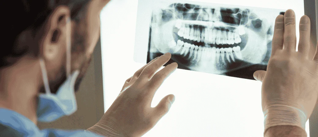
What Periapical and Panoramic X-Rays Can Show
Standard dental imaging is the first step. Radiographic evaluation plays a central role in spotting resorption and judging how far it has spread.
A periapical radiograph shows one or two teeth from crown to root tip. It’s the go-to image for checking root length and looking for dark areas inside the root.
A panoramic radiograph (also called an OPG) captures all your teeth and both jaws in one wide image. It’s useful for screening, though fine details can be harder to read.
In fact, many cases of internal resorption are found on X-rays taken during routine check-ups, not because a patient complained.
When CBCT Provides a Clearer Three-Dimensional View
Flat images have limits. Two roots can overlap, and a defect on the tongue side of a tooth may hide behind healthy structure.
That’s when your dentist may order a cone beam computed tomography (CBCT) scan. CBCT imaging builds a three-dimensional picture, so the lesion can be viewed from several angles.
Early diagnosis with X-ray or CBCT gives the best chance of keeping the tooth. A scan helps your dentist measure how deep the defect goes, whether it reaches the pulp, and if root canal treatment or extraction makes more sense.
CBCT is also helpful for telling different types of resorption apart, such as internal versus external cervical resorption.
Balancing Diagnostic Detail With Radiation Safety
More detail isn’t always better. Dentists follow the idea of using the lowest dose that still answers the question.
Digital sensors need far less radiation than old film. Lead aprons, thyroid collars, and tight beam settings lower your exposure further.
Your dentist decides how often to image based on your risk, not a fixed calendar. Past trauma, orthodontic treatment, or a history of resorption may mean more frequent checks.
CBCT delivers a higher dose than a single periapical film, so it’s ordered when it will change your treatment plan.
Even so, these images reveal problems a visual exam can’t detect, which is why skipping them isn’t the safer choice.
Treatment, Monitoring, and Long-Term Tooth Preservation
Once your dentist spots resorption on an X-ray, the next step depends on where the damage sits, how fast it is moving, and how much healthy root is left.
Some teeth only need watching, others need endodontic treatment or surgery, and a few are better off removed and replaced.
| Situation | Typical approach |
| Small, stable external spot | Watch with repeat X-rays |
| Internal resorption with infected or necrotic pulp | Root canal therapy |
| Defect that can’t be reached through the canal | Surgical repair or apicoectomy |
| Perforated root, loose tooth, little root left | Extraction and replacement |
When Monitoring Is Appropriate for Small Stable Lesions
Not every spot needs immediate treatment. If the lesion is tiny, the pulp still responds normally, and there’s no pain or swelling, your dentist may simply keep an eye on it.
Monitoring usually means a follow-up X-ray in 6 to 12 months, since radiographic evaluation of root resorption is the main way to tell if a defect is growing. A cone beam CT scan may be added if flat films look unclear.
If the cause was pressure from braces or an impacted tooth, removing that pressure often stops the process.
Keep in mind that lost root structure does not grow back, resorption is irreversible, so the goal of watching is to catch change early, not to wait for symptoms.
Root Canal Therapy for Internal or Infection-Related Damage
Internal resorption starts inside the canal, and it feeds off inflamed or dying pulp tissue. That means a root canal is usually the fix.
Your endodontist opens the tooth, cleans the canal, and works to remove all vital and necrotic tissue so the resorbing cells lose their fuel.
Because resorption creates odd, bumpy spaces that files can’t reach, extra steps help, ultrasonic irrigation with sodium hypochlorite, plus a calcium hydroxide dressing left in place for two to four weeks.
At the filling appointment, warm gutta-percha is often used instead of standard cones because it flows into irregular shapes better.
Done early, root canal treatment can stop internal resorption and keep the tooth working for years.
Surgical Repair and Apicoectomy for Selected Cases
Some defects sit on the outside of the root, near the gumline, where a file can never reach. Those cases call for surgical intervention.
Your endodontist lifts a small gum flap, cleans out the soft, discolored tissue, and seals the cavity with a calcium silicate material like MTA or Biodentine.
The area may also be filled with bone graft granules to help support the root.
An apicoectomy is used when damage is limited to the root tip. The surgeon removes the last few millimeters of root and seals the end.
These procedures are more predictable when the defect is caught before it invades deeply. Advanced cervical resorption that wraps around the root has a poorer outlook.
When Tooth Extraction and Replacement Are Needed
Sometimes saving the tooth just isn’t realistic. Extraction is usually recommended when the root is perforated in several places, when tooth mobility is grade 2 or higher, or when so little root remains that a crown could never be supported.
Teeth with replacement resorption, where bone slowly takes over the root, also can’t be repaired.
The good news is that prosthetic replacement options are solid. A dental implant, a fixed bridge, or a removable partial can restore chewing and appearance.
Ask your dentist about the likelihood of saving the tooth, expected treatment costs, and its long-term outlook before deciding how to proceed.
Protecting Teeth After Treatment
After a repair, a few habits go a long way.
- Keep up regular dental check-ups. Yearly X-rays catch new lesions on other teeth before they cause trouble.
- Wear a mouthguard for contact sports and a night guard if you grind. Trauma is a leading trigger.
- Brush and floss carefully around repaired areas, since good oral hygiene keeps gum inflammation from adding stress to the root.
- Speak up during orthodontics. Light forces and periodic X-rays lower risk, because the inflammation needed to move a tooth is also what drives resorption.
- Report new pain, a pink spot on the crown, or looseness right away.
Frequently Asked Questions
Root resorption breaks down tooth structure from the inside or outside, often without any pain to warn you. Below you’ll find clear answers about how it’s spotted on X-rays, what triggers it, and which treatments can stop it.
What is root resorption, and how serious is it?
Root resorption happens when your own cells start dissolving the hard tissue of a tooth root. The immune system dissolves the tooth root structure, which can leave the tooth loose or cause it to fall out.
It’s a normal process in kids losing baby teeth. In adults, though, it points to a dental problem that needs attention.
How serious it gets depends on timing. A small, stable spot may just need watching, while advanced damage can mean losing the tooth.
Two cell types do the work. Odontoclasts break down dentin, and osteoclasts break down cementum and bone.
Does root resorption show up on routine dental X-rays?
Yes, and that’s usually how it’s found. Most cases show up by chance on X-rays taken during a regular checkup.
Your dentist may see a dark shadow in or around the root. Since resorption rarely hurts early on, these images often give the first clue.
Sometimes a standard X-ray isn’t enough. A 3D scan called a CBCT gives a clearer picture, and advanced imaging like CBCT helps pinpoint the exact type and size of the defect.
That’s one more reason to keep up with your checkups. Waiting years between visits gives resorption time to spread quietly.
What causes internal and external root resorption?
Both types start when the tooth’s protective layers get damaged. Cementum covers the outside of the root, and a layer called predentin lines the pulp chamber inside.
Internal resorption begins inside the root canal or pulp chamber. It’s often linked to long-term inflammation or infection in the pulp, sometimes after an injury.
External resorption starts on the outer root surface. Common triggers include:
- A blow to the tooth or other dental trauma
- Pressure from braces or aligners
- Infection around the root tip
- Pressure from an impacted tooth or a cyst
- Certain health conditions affecting bone
Braces are a known factor, and some tooth movement causes minor resorption in a share of orthodontic patients. Most of those cases stay mild and don’t threaten the tooth.
What symptoms can indicate root resorption?
Early on, you probably won’t feel anything at all. That’s why it’s often called silent.
As it advances, you might notice:
- Tooth sensitivity or a dull ache
- A pink or dark tint showing through the enamel
- A tooth that feels loose
- Gum swelling or tenderness near one tooth
- A tooth that shifts position or looks shorter
- Pain when you bite down
Any of these deserve a visit to your dentist. Early diagnosis can stop further damage and may keep you from losing the tooth.
How quickly can root resorption progress?
There’s no single timeline. Some cases creep along slowly for years, while others move much faster.
Speed depends on the cause. Resorption driven by an active infection tends to advance more quickly than mild damage from orthodontic pressure, which often stops once the braces come off.
Because the pace varies so much, your dentist may take follow-up X-rays a few months apart. Comparing images over time shows whether the area is holding steady or growing.
What treatment options are available for root resorption?
Treatment depends on the type, the location, and how much tooth structure is left. The lost tissue can’t grow back, but treatment can halt its progression and save the tooth when it’s caught early.
Common approaches include:
- Root canal treatment to clear out infected pulp and stop internal resorption
- Surgical repair to clean the damaged spot on the outer root and seal it with a filling material
- Removing the trigger, such as pausing orthodontic treatment or taking out an impacted tooth
- Monitoring with periodic X-rays for small, stable areas
- Extraction when too much root has been lost, followed by an implant or bridge
Catching it sooner gives you more choices. Depending on the cause and how early it’s found, treatment may stop or slow the process and save the tooth.


